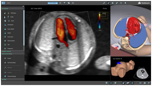
Fetal echocardiography is a test that uses sound waves (ultrasound) to evaluate the baby's heart for problems before birth.
Fetal echocardiography is a test that is done while the baby is still in the womb. It is most often done during the second trimester of pregnancy. This is when a woman is about 18 to 24 weeks pregnantv.
The procedure is similar to that of a pregnancy ultrasound. You will lie down for the procedure. The test can be performed on your belly (abdominal ultrasound) or through your vagina (transvaginal ultrasound).
In an abdominal ultrasound, the person performing the test places a clear, water-based gel on your belly. A hand-held probe is moved over the area. The probe sends out sound waves, which bounce off the baby's heart and create a picture of the heart on a computer screen.
In a transvaginal ultrasound, a much smaller probe is placed into the vagina. A transvaginal ultrasound can be done earlier in the pregnancy and produces a clearer image than an abdominal ultrasound.

This test is done to detect a heart problem before the baby is born. It can provide a more detailed image of the baby's heart than a regular pregnancy ultrasound.
The test can show:
The test may be done if: